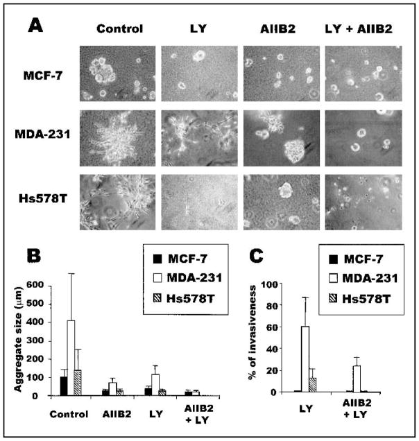
Fig. 1.
Inhibiting the combination of β1 integrin and phosphatidylinositol 3-kinase (PI3K) promotes a greater alteration in morphology, aggregate size, and invasiveness than inhibiting either PI3K or β1 integrin alone. A) MCF7, MDA-MB-231 (MDA-231), and Hs578T breast tumor cells grown in three-dimensional (3D) reconstituted basement membrane (rBM) cultures in the presence of anti-β1 integrin antibody (AIIB2) and/or PI3K inhibitor LY294002 (LY). All cultures were analyzed after 10 days of rBM culture. B) Size of the colonies formed by the three breast tumor cell lines grown in 3D rBM in the presence of inhibitors (error bars indicate 95% confidence intervals of triplicate samples). Experiments were repeated four times with similar results. C) Invasiveness of treated and untreated MDA-MB-231 (MDA-231) and Hs578T breast cancer cells in Matrigel-coated Boyden chamber assays. The invasiveness of these cells treated with AIIB2, LY294002, or AIIB2 plus LY294002 is shown as a percentage of control (error bars indicate 95% confidence intervals of triplicate samples; experiments were repeated four times with similar results). MCF7 cells were not invasive in this assay. Experiments were repeated three times with similar results.

