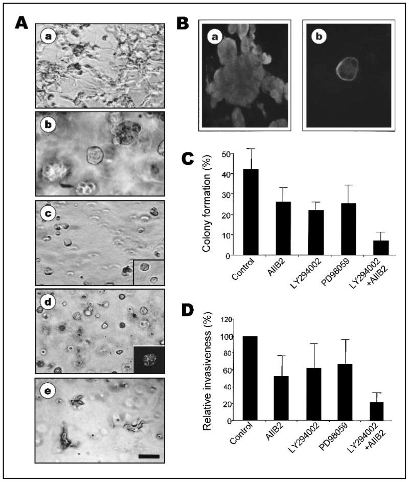
Fig. 3.
Inhibition of β1 integrin plus phosphatidylinositol 3-kinase (PI3K) or inhibition of β1 integrin plus mitogen-activated protein kinase (MAPK) is sufficient to induce phenotypic reversion of MDA-MB-231 cells. A) Phase-contrast micrographs of control untreated MDA-MB-231 cells (a), MDA-MB-231 cells treated with β1 integrin inhibitory antibody AIIB2 (b), AIIB2 plus PI3K inhibitor LY294002 (c), AIIB2 plus MAPK inhibitor PD98059 (d), or LY294002 plus PD98059 (e) in three-dimensional (3D) reconstituted basement membrane (rBM) cultures. Bar = 50 μm. c, inset) Phase-contrast micrograph of S1 cells in 3D rBM. d, inset) Confocal fluorescence microscopy image of filamentous actin (F-actin) in 5-μm cryosections of MDA-MB-231 cells treated with AIIB2 plus PD98059 (×2 image). Bar = 25 μm. Similar pattern of staining of F-actin and nuclei was obtained with cells treated with AIIB2 plus LY294002. B) Confocal immunofluorescence microscopy images of β4 integrin in untreated control (a) and AIIB2 plus LY294002 treated (b) MDA-MB-231 cells. C) Anchorage-independent growth of untreated and treated MDA-MB-231 cells in soft agar. Colony formation by MDA-MB-231 cells treated with AIIB2, LY294002, PD98059, or AIIB2 plus LY294002 shown as percentage of colony formation (mean ± 95% confidence intervals; experiments were repeated three times with similar results). D) Relative invasiveness of untreated and treated MDA-MB-231 cells, treated as in (B), shown as percentage of control (± 95% confidence intervals; experiments were repeated three times with similar results).

