
Figure 6.
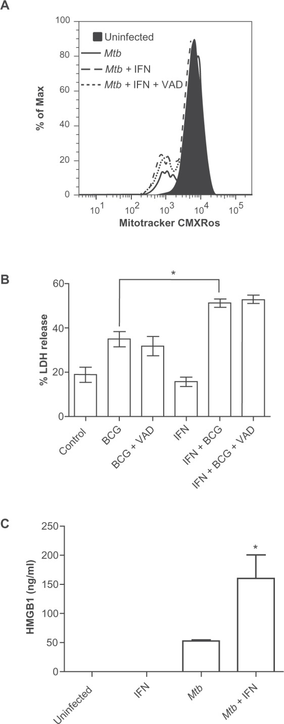
IFN-γ increases necrosis heavily infected macrophages as determined by loss of ΔΨm A) increased LDH release B) and increased HMGB1 release C) from heavily infected macrophages. A) Cells infected with Mtb Erdman at MOI of 25 for 3 h were stained with MitoTracker CMXRos to measure ΔΨm. The filled peak corresponds to uninfected cells, the solid line corresponds to Mtb-infected cells, while the dashed and dotted lines correspond to Mtb-infected cells treated with IFN-γ or IFN-γ plus Z-VAD-fmk, respectively. Histograms are representative of three independent experiments. B) For LDH assays IFN-γ pretreated BMDM were infected with BCG at MOI of 25 for 9 h and then supernatant was collected for measurement of LDH. Data are means ± SEM of four independent experiments. C) For HMGB1 assays cells were infected with Mtb Erdman at MOI of 25 for 6 h. Culture supernatants were collected and HMGB1 was measured by antigen-capture ELISA. Data are presented as the mean ± SD from two independent experiments. *p < 0.05.