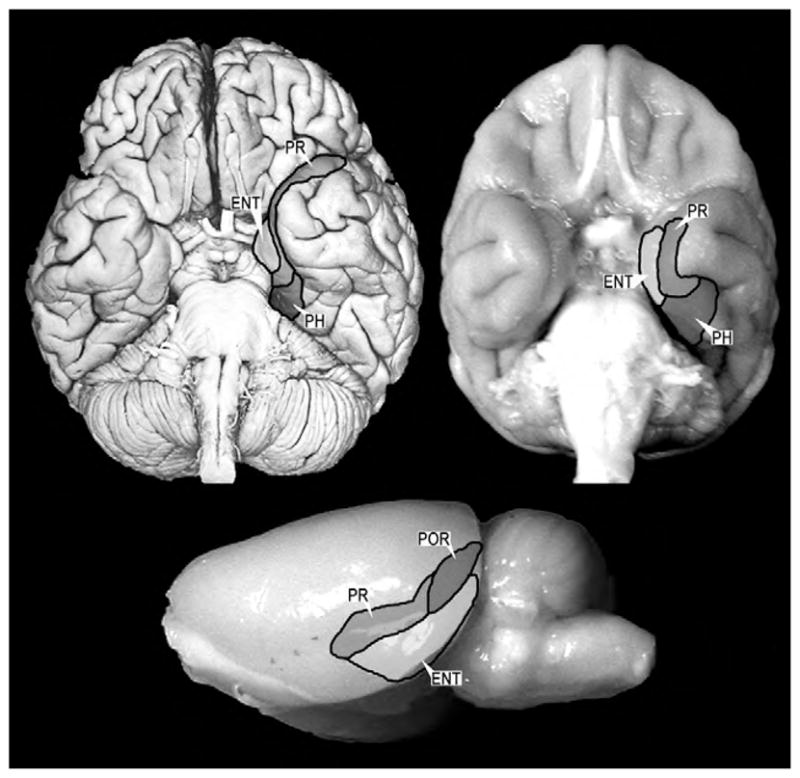
Fig. 1.

Ventral view of a human brain (upper left), a monkey brain (upper right) and a lateral view of a rat brain (lower center). The major cortical components of the medial temporal lobe are highlighted and outlined. The organization and connections of these structures are highly conserved across these species. Abbreviations: PR: perirhinal cortex, PH: parahippocampal cortex, ENT: entorhinal cortex, POR: postrhinal cortex (referred to as parahippocampal cortex in primates).
