
Figure 2.
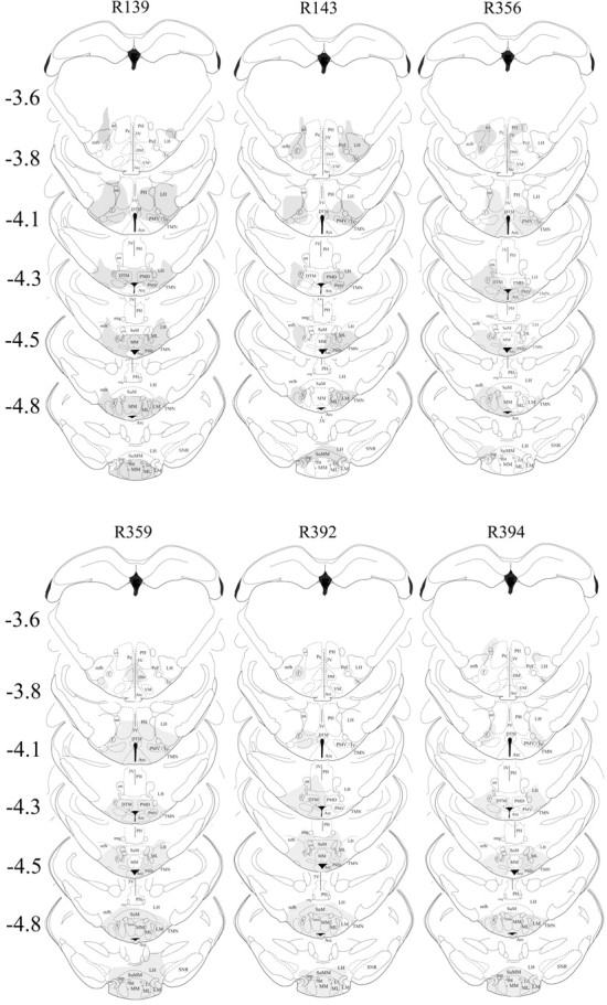
Extent of neuronal loss because of local injection of HCRT2-SAP (250 ng/μl in 0.25 μl each side) into the TMN. HCRT2-SAP kills neurons that possess the HCRT-2 receptor, which is on the histaminergic TMN neurons but also on other adjacent neurons. Thus, the neuronal marker NeuN was used to assess the extent of nonspecific neuronal loss, and those regions devoid of neurons were then drawn onto the corresponding figures taken from the rat brain atlas (Paxinos and Watson, 2006). Each column represents an individual rat (marked by number at the top of each column) in the triple-lesion group (BF+TMN+LC). The numbers to the left indicate anteroposterior distance from bregma in millimeters. Arc, Arcuate n; DM, dorsomedial hypothalamus; DTM, dorsal tuberomammillary n; MM, medial mammillary n; MMn, median mammillary n; ML, medial mammillary n lateral; LH, lateral hypothalamus; LM, lateral mammillary n; PeF, perifornical area; PH, posterior hypothalamus; PMD, premammillary n dorsal; SuM, supramammillary n; SNR, substantia nigra reticular; VM, ventromedial hypothalamus; Te, Terete hypothalamic n; 3V, third ventricle; f, fornix; mfb, medial forebrain bundle; mt, mammilothalamic tract; mtg, mammilotegmental tract; mp, mammillary peduncle.