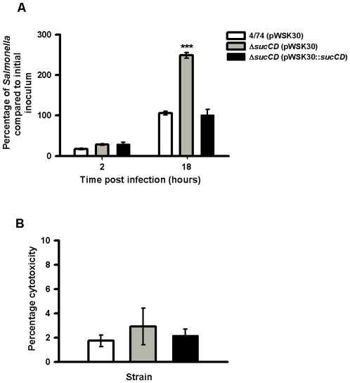
Figure 3. Complementation of the S. Typhimurium ΔsucAB strain in resting RAW macrophages and cytotoxicity assays.
(A) Numbers of viable bacteria (expressed as percentages of the initial inoculum) inside the macrophages at 2 h and 18 h after infection. Each bar indicates the statistical mean for three biological replicates, and the error bars indicate the standard deviations. The significant differences between the parental 4/74 strain and the mutant and complemented strains are shown by asterisks p>0.05, * p<0.05, ** p<0.01, and *** p<0.001. (B) Cytotoxicity assays of S. Typhimurium wild-type, ΔsucCD (AT3449) and complemented ΔsucCD (AT???) strains in RAW macrophages after 18 h infection as a percentage of total LDH release from lysed uninfected macrophages. All cytotoxicity data were obtained from three independent biological replicates.

