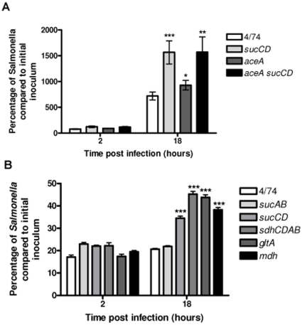
Figure 4. Increased intracellular replication of the S. Typhimurium ΔsucCD, ΔsdhCDAB, ΔgltA and Δmdh strains during infection of activated RAW macrophages.
(A) Intracellular replication assays of S. Typhimurium 4/74, ΔsucCD (AT3449), ΔaceA (AT3385) and ΔaceAΔsucCD (AT3496) strains during infection of resting RAW macrophages (B) Intracellular replication assay of S. Typhimurium 4/74, ΔsucAB (AT3448), ΔsucCD, (AT3449), ΔsdhCDAB (AT3475), ΔgltA (AT3505) and Δmdh (AT3508) strains during infection of activated RAW macrophages The data show the number of viable bacteria (expressed as percentages of the initial inocula) within activated macrophages at 2 h and 18 h post-infection. Each bar represent the statistical mean from three independent biological replicates and the error bars represent the standard deviation (The significant differences between the parental 4/74 strain and the mutant strains are shown by asterisks p>0.05, * p<0.05, ** p<0.01, and *** p<0.001).

