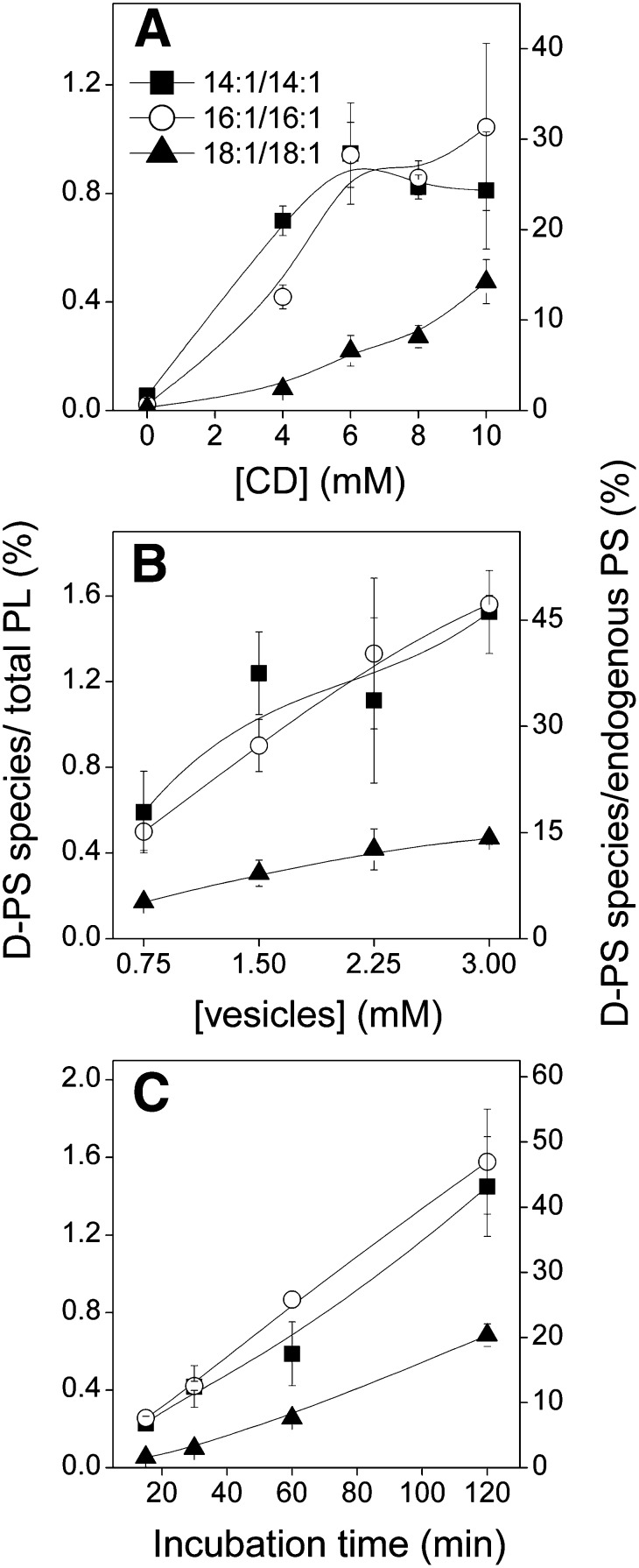
Fig. 1.
Effect of meβ-CD or donor vesicle concentration and incubation time on cellular uptake of exogenous PS. A: BHK cells were incubated for 1 h with 1.5 mM donor vesicles containing cholesterol, POPC, and labeled 14:1/14:1- (squares), 16:1/16:1- (circles), and 18:1/18:1-PS (triangles) (30:27:1:1:1, mol/mol) and indicated concentration of meβ-CD. B: BHK cells were incubated for 1 h in the presence of 8 mM meβ-CD and indicated concentration of the vesicles described in A. C: BHK cells were incubated with vesicles and meβ-CD as in A for the indicated times. In each experiment, 1.5 mM hydroxylamine and 25 μM MAFP were included to prevent decarboxylation and inhibit acyl chain remodeling of exogenous PS, respectively. Symbols as indicated in A. The amount of PS incorporated is shown as the percentage of the total phospholipid (left Y-axis) or relative to endogenous PS (right Y-axis). Data are means of three (A) and two (B and C) independent experiments ± SD.

