
Fig. 7.
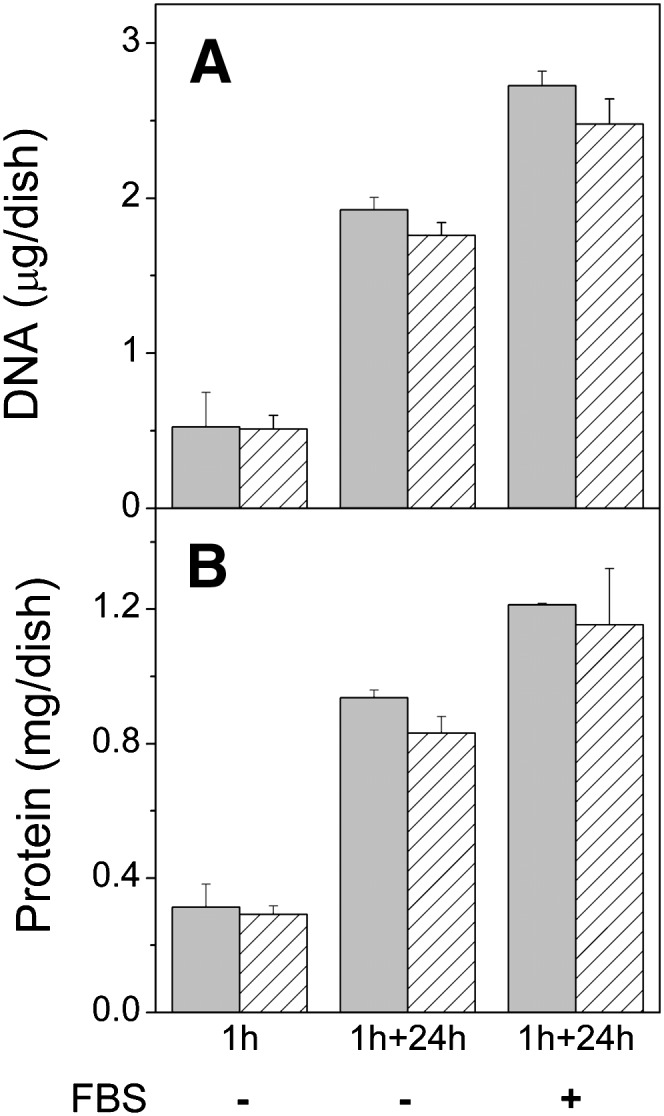
Effect of incubation with vesicles and meβ-CD on cell growth. BHK21 cells were incubated for 1 h in the presence of 8 mM meβ-CD and 0.5 mM donor vesicles composed of cholesterol, POPC, and 14:1/14:1-PS (10:9:1 mol/mol). Cells were then washed and chased in DMEM for 0 h or 24 h in absence (-) or presence (+) of 10% FBS. DNA (A) and protein (B) contents of the cells were then determined. Gray bars are cells incubated with vesicles and meβ-CD, striped bars, untreated cells. Data are means of three parallel samples ± SD. Parallel results were obtained in experiments with somewhat different incubation and chase times.