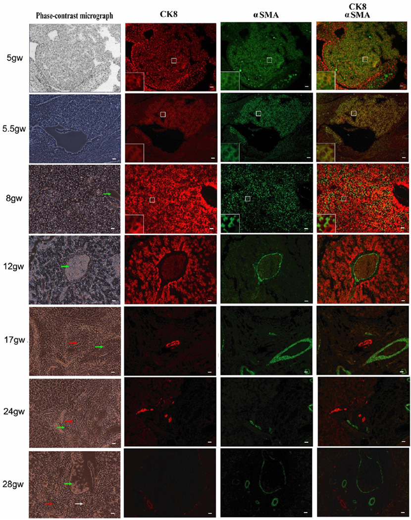
Fig. 1. Immunofluorescent study of human fetal livers from 5gw to 28gw.
Multi-immunostaining with R555-conjugated anti–CK8 (red), FITC-conjugated anti-αSMA (green) and DAPI (blue). Co-expression of CK8 and αSMA is observed in most non-hematopoietic cells of human fetal liver at early gestational stages (5gw and 5.5gw), in only a few cells at 8gw, but in none of the cells at later gestational stages (12gw, 17gw, 24gw and 28gw). By 12gw, expression of CK8 and αSMA was segregated. Some cells in centre box were magnified as shown in left-down box. Arrows in green, white and red represent interlobular veins, interlobular arteries and bile ducts respectively. Scale bar = 20 µm.

