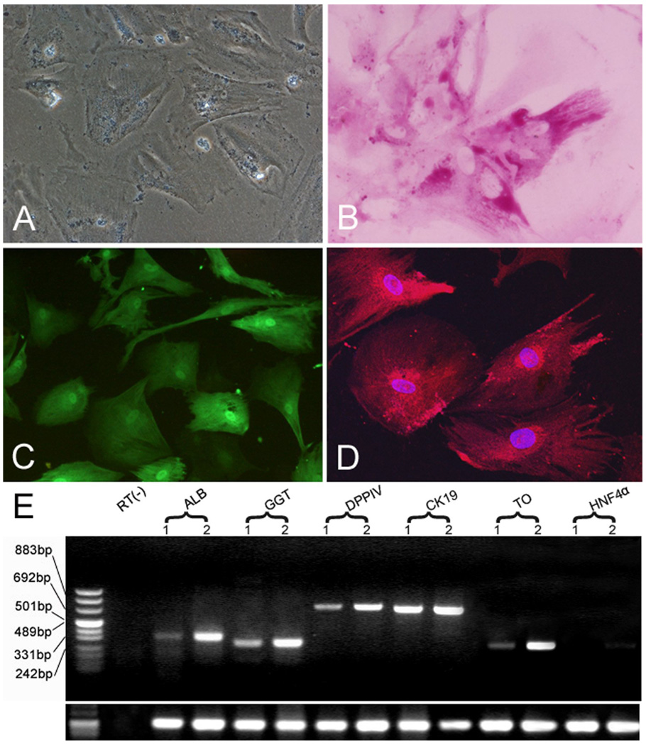
Fig. 4. Hepatic differentiation of hLBSC.
Binuclear cells were observed in 2 days after SB treatment, and by 10 days, binuclear cells comprised of 10–15% of the total cell population (A). Abundant glycogen stores were seen in the cytoplasm of most binuclear cells by PAS staining (B). Immunocytofluorescene showed expression of TAT and ALB in the hLBSC treated by SB (C, D). Enhanced expression of ALB, GGT DPPIV, CK19, TO and HNF4α was detected by RT-PCR (E). Lane 1: samples without SB treatment; Lane 2: samples with SB treatment. RT (−): mRNA sample without reverse transcription.

