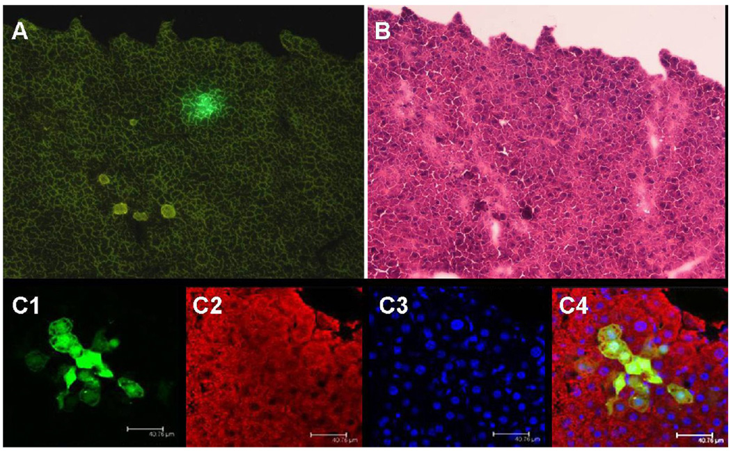
Fig. 6. In vivo transplantation of hLBSC into CCl4-treated SCID mice.
hLBSC- pLNCG C1 cell line with stable EGFP expression was established. Then the EGFP expressing cells were transplanted introsplenicly into SCID mice pre-treated by CCL4. Three weeks after implantation, EGFP-positive cell clusters were detected in the recipient mice (A). H&E staining confirmed that they were clusters of hepatocytes in the hepatic cord (B). The overlay image of EGFP (green, C1) and the same section stained by anti-α-1-AT antibody (red, C2) clearly shows that all EGFP-positive donor cells were also α-1 AT-positive hepatocytes (yellow, C4), with DAPI nuclear counterstaining (blue, C3). (Relative magnification A, B: 100×; C: 200×)

