
Figure 6.
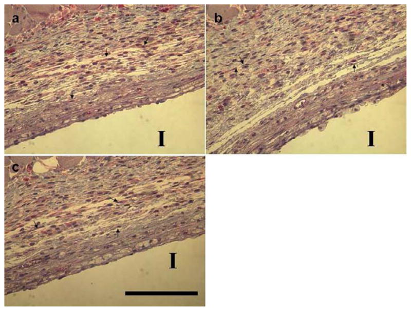
Immunostains of mTOR protein expression in foreign body capsules from murine histological sections. Tissue samples surrounding implants (I) were harvested from mice two-week post-implantation. Immunohistochemical staining for mTOR in foreign body capsules around filter paper from (a) negative control group (no siRNA loaded), (b) 2μg mTOR siRNA-treated group, and (c) 10μg mTOR siRNA-treated group. Sections were stained with mTOR antibody, and counterstaining was done with hematoxylin. These treatments stain target proteins red and cell nuclei dark (blue). Arrows denote fibroblasts (scale bar = 125μm).