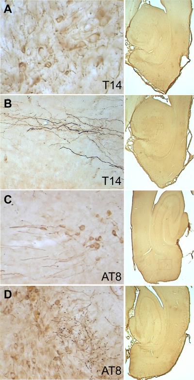
Figure 2.
Human tau immunoreactivity was distributed throughout all neuronal cytoplasmic compartments including perikarya, dendrites (panel A, entorhinal cortex), and axons (panel B, presumptive perforant path projections in dentate gyrus). Hyperphosphorylated tau was similarly localized in somata, dendrites (panel C, entorhinal cortex), axons and synaptic boutons (panel D, presubiculum). Controls were blank for human tau immunoreactivity. Boxes in the 1× images at right show the approximate location of each higher-magnification frame.

