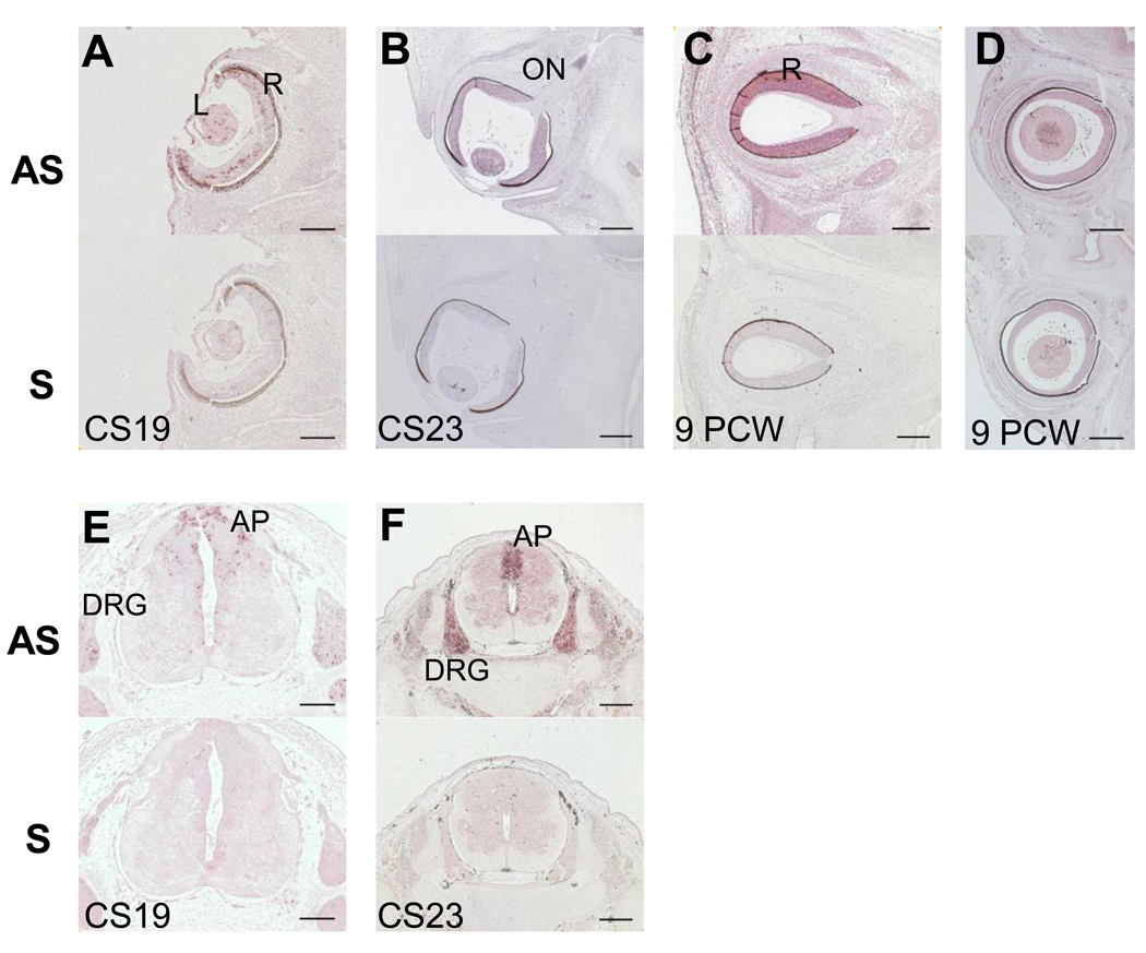
Figure 2.
PLA2G6 expression in human eye and spinal cord at CS19 (A,E) CS23 (B, F) and 9PCW (C,D). Each panel has 2 images, the top one showing in situ hybridization using antisense (AS) probes and the lower one using sense (S) probes for PLA2G6. No signal was detected using sense control probes A) PLA2G6 staining in retina and lens at CS19. B) PLA2G6 staining in lens, retina and optic nerve at CS23. C–D) PLA2G6 staining in retina at PCW9. E) PLA2G6 staining in dorsal root ganglia at CS19. F) PLA2G6 staining in dorsal root ganglia at 9PCW A- alar plate, AS- antisense, DRG- dorsal root ganglia, L- lens, ON- optic nerve, R-retina, S-sense, Scale bars are: 100µm in A,E; 200µm in B,C,D,F.

