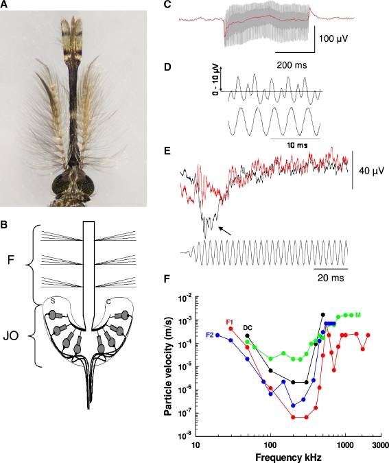
FIG. 1.
Electrical potentials recorded from the Johnston’s organ and thorax of mosquitoes. A Head and antennae of male A. gambiae mosquito with fibrillae extended. B Schematic diagram showing a cross-section of the antenna of a mosquito with the flagellum of the antenna (F) inserted into the cup-shaped pedicel that houses the complex arrangement of cuticular processes (C) and attached, mechanosensory scolopidia (S) of the Johnston’s organ (JO; Belton 1989). C Receptor potential (gray) with direct current (DC) component (red line) from a male A. gambiae with collapsed fibrillae in response to a 300 Hz tone, particle velocity 0.0011 ms−1. D Extracellular (double frequency) receptor potential recordings from the JO of a male Culex pipiens in response to a 300.3 Hz, 36 dB SPL, 0.00405 ms−1 tone (command voltage to speaker, lower trace; modified from Pennetier et al. 2010, with permission of the publisher). E Compound voltage responses (upper trace) recorded from the JO of male M-form A. gambiae mosquito in response to a 300.3 Hz tone, particle velocity 0.56 mm s−1 (lower trace) before (black) and after (red) injecting 1 μM tetrodotoxin (TTX, Sigma-Aldrich) in insect saline into the thorax. Note loss of onset (neural) potential, but not phasic (2f) component after TTX injection. F Threshold frequency tuning curves measured from of the F1, F2, and DC components of the extracellular receptor potential recorded from the JO of a male C. pipiens and neural motor (M) responses recorded from the thorax (E and F modified from Warren et al. 2010, with permission of publisher (all Anopheles figures modified from Warren et al. 2009, with permission of publisher).

