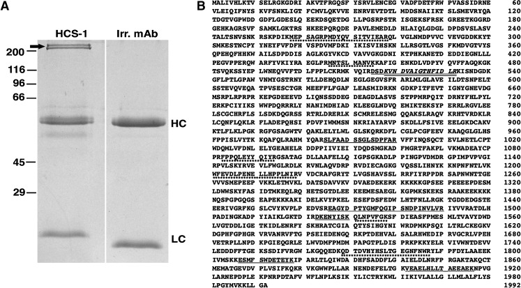
FIG. 2.
Identification of HCS-1 antigen as otoferlin. A Immunoprecipitation from chick sensory epithelia using the HCS-1 antibody. Coomassie-stained SDS-PAGE gel revealing a high molecular weight doublet immunoprecipitated from a Triton X-100 extract of chicken inner ear (arrow, left lane). These proteins were not immunoprecipitated with the H27 antibody, an isotype-matched irrelevant antibody (right lane). Immunoprecipitated bands were excised and subjected to mass spectrometry. Bands corresponding to the immunoglobulin heavy- and light chains are indicated by HC and LC, respectively. Molecular mass markers in kilodaltons are indicated to the left. B Location of tryptic peptides in the sequence of mouse cochlear otoferlin. Tryptic digestion followed by mass spectrometry identified seven peptides unique to the otoferlin sequence for the 230-kDa immunoprecipitated protein (underlined). One of these peptides (italics) overlapped a larger peptide. The 210-kDa immunoprecipitated protein identified the six of the same peptides obtained for the higher molecular weight band and an additional six peptides (underlined with asterisks).

