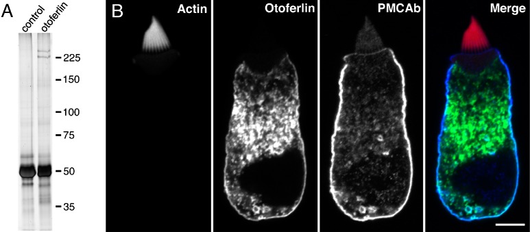
FIG. 8.
Immunoprecipitation of otoferlin from bullfrog saccule and subcellular localization in isolated bullfrog saccular hair cells. A Silver stain of a SDS-PAGE gel containing proteins immunoprecipitated from bullfrog sacculus with the anti-otoferlin antibody (HCS-1) or an irrelevant isotype-matched control antibody (anti-V5 antibody). Immunoglobulin heavy chain is observed at approximately 50 kDa. Molecular mass markers in kilodaltons are indicated to the right. B Confocal localization of actin (red), otoferlin (HCS-1 antibody, green), and PMCAb (blue) in an isolated saccular hair cell by confocal microscopy. Individual channels are shown to the left, merged image is shown to the right. Scale bar = 5 μm.

