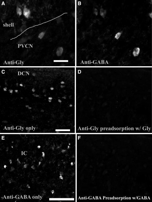
FIG. 1.
Immunoreactivity of glycine and GABA in the CN. a and b Photomicrographs of glycine and GABA immunoreactivity in the VCN (40×). Both photomicrographs (a and b) are taken from the same CN region. The line in a indicates the boundary between the shell region and central PVCN. The two large neurons shown in central VCN are clearly colabeled with both glycine (a) and GABA (b). The two shell neurons are also colabeled but very lightly for GABA. c Photomicrographs of glycine immunoreactivity in DCN layer 2 (20×). Cells show positive glycine immunoreactivity and likely are cartwheel cells. d Preadsorbtion of antiglycine antibody with glycine-BSA resulted in negative immunoreactivity in DCN layer 2. e Photomicrographs of immunoreactivity of GABA in the IC (20×). f Preadsorbtion of anti-GABA antibody with GABA-BSA resulted in negative immunoreactivity in IC. Scale bars in a, c, e = 50 µm.

