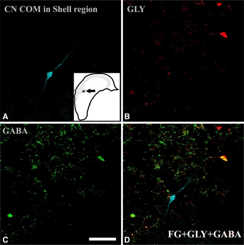
FIG. 3.
Confocal images (40×) of a retrogradely labeled CN-commissural neuron in the shell region of PVCN showing negative glycine immunoreactivity and negative GABA immunoreactivity. a The retrogradely labeled cell in the shell region following injections of FG in the contralateral CN. The inset shows the location of the labeled cell. This cell shows both negative glycine immunoreactivity (b) and negative GABA immunoreactivity (c). Confocal multichannel image (overlap of a, b, and c) shows no colocalization of FG with either glycine or GABA immunoreactivity (d). Scale bar = 50 µm.

