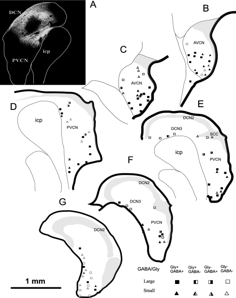
FIG. 5.
Schematic drawing of CN-commissural neurons with their glycine and GABA immunoreactivity. a The injection site in the contralateral CN. b–g Drawings of CN transverse sections that evenly extend from the caudal to rostral ends of the right CN. The labeled CN-commissural neurons were manually counted on every fourth section and mapped on the corresponding templates. Each symbol represents a labeled neuron. Symbols that represent immunoreactivity to glycine or GABA are illustrated in the right lower corner. The majority of glycine negative (−) and GABA negative (−) CN commissural neurons are located in the medial and shell regions of VCN. Scale bar = 50 μm. DCN2 fusiform cell layer also containing granule cells and therefore part of GCD, DCN3 deep layer of DCN.

