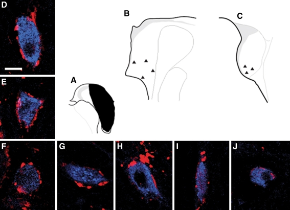
FIG. 8.
Confocal images showing retrogradely labeled commissural neurons with somata surrounded by VGLUT2 immunoreactive terminals. A Schematic FG injection site in contralateral CN. B Schematic of the locations of commissural neurons in G–J. These neurons are located in the ventral part of AVCN, and have oval or round or elongate cell bodies with medium to large size. c Schematic of the locations of the commissural neurons in D–F. These neurons are located in the ventral part of PVCN, have oval or round large cell bodies, and are surrounded by positive VGLUT2 immunoreactive terminals.

