Figure 7.
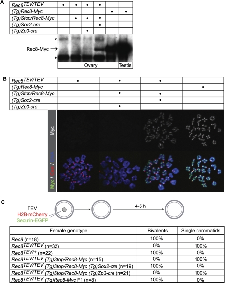
No cohesin turnover in growing-phase oocytes. (A) Western blot analysis of Rec8-Myc using c-Myc antibody on whole ovary and testis extracts. Rec8-Myc is expressed constitutively from either a multicopy (Tg)Rec8-Myc BAC transgene (second row) or a (Tg)Stop/Rec8-Myc BAC transgene carrying a conditional Stop cassette flanked by LoxP sites (third row) that is activated by Cre recombinase under the control of the Sox2 (fourth row) or Zp3 (fifth row) promoter. The asterisks indicate cross-reacting bands with c-Myc antibody. (B) Chromosome spreads were prepared from four types of oocytes at 5 h post-GVBD. Chromosome spreads were stained with c-Myc antibody to visualize Rec8-Myc (green), CREST to mark centromeres (red), and Hoechst to visualize DNA (blue). (C) Seven types of GV oocytes expressing TEV protease, H2B-mCherry, and Securin-EGFP were cultured in the presence of IBMX for 1 h, then released into IBMX-free culture medium, and chromosomes were visualized by time-lapse confocal microscopy. Oocytes (n = number of cells) were scored as containing either 20 bivalent chromosomes or at least 72 single chromatids (and no bivalents) by 5 h post-GVBD.

