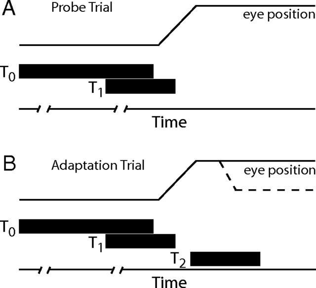
Figure 1.
Schematic representation of trial types. The delayed probe trial began with the presentation of the visual target at T0 (A). After a variable fixation interval (indicated by line breaks along the time axis) a second target was presented. This target could be at the T1 location or at other locations used to define the movement field of rerecorded cells. Subjects were not permitted to look to the T1 target until target T0 was turned off. Saccade initiation was detected using a position criterion and T1 was then turned off. The delayed adaptation trials were identical until 20–40 ms after T1 was turned off. T2 was now illuminated and remained lit until the subject fixated this location for 500 ms. Dotted lines in B indicate the corrective saccade that to the T2 location during backward adaptation.

