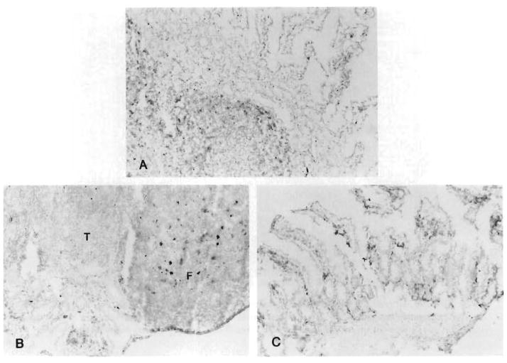
Fig. 8.
Replacement of the donor lymphoid tissue with recipient cells. A, Twelve days after MVTX under high-dose FK 506 for the 12 days of survival (group 3) about one half of the intestinal lymphoid cells were replaced (L-21-6, red; hematoxylin hematoxyling counterstain). B, At 114 days (group 3), most of the cells in the follicles of the Peyer’s patches displayed recipient Ia determinants. C, Same replacement in the lamina propria of the villi (group 3) at 134 days. (L-21-6, red; hematoxylin counterstain; original counterstain (original magnification X300.) T, T-cell interfollicular zone; F, B-cell follicle.

