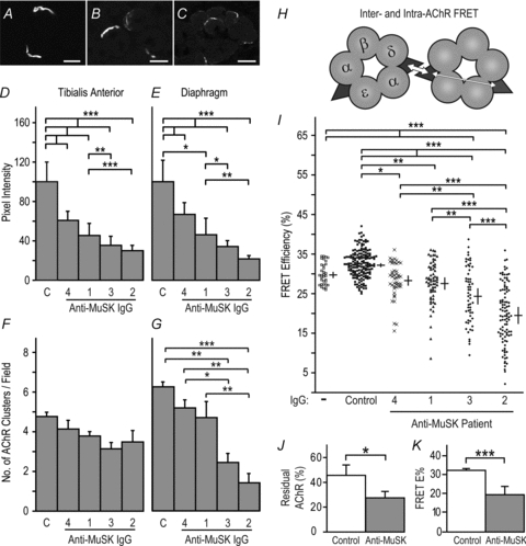
Figure 1. MuSK IgG reduced postsynaptic AChR packing density.

Mice were injected with human IgG (45 mg day−1, i.p.) for 14 days before killing. A, transverse optical section showing bright postsynaptic AChR plaques at NMJs in the TA muscle of a mouse injected with control human IgG. B and C, reduced postsynaptic AChR staining in mice injected with MuSK3 or MuSK2 IgGs, respectively. D and E, intensity of postsynaptic AChR staining at synapses in the TA and diaphragm muscles, respectively. The source of the IgG (patients 1–4 or control, C) is indicated beneath each bar. F and G, counts of the number of AChR plaques (NMJs) per photomicrograph field in the TA and diaphragm muscles, respectively. H, fluorescently labelled α-bungarotoxin molecules bound to AChRs function as donor and acceptor fluorophors (filled triangles) for FRET. Inter-AChR FRET (thick arrow) is much more efficient than intra-AChR FRET (thin arrow; ∼30% cf. ∼5%) because FRET efficiency declines sharply with distance (Gervásio et al. 2007). I, efficiency of postsynaptic AChR-AChR FRET. Symbols show the range of FRET efficiencies for individual synapses in TA muscles. Crossed bars show the mean endplate FRET efficiencies for the 3 mice in each treatment group (±s.e.m.). J, percentage of postsynaptic fluorescent α-bungarotoxin staining intensity remaining after extracting cryosections with non-ionic detergent. Mice injected with MuSK2 IgG for 14 days showed a significant reduction in the fraction of AChR that was resistant to detergent extraction (residual AChR), compared to control mice. K, FRET data for MuSK2 IgG-injected mice re-plotted from panel I for comparison. Data in panels D–G and I–K represent mean +s.e.m. for n= 3 mice. Scale bars represent 30 μm for panels A and B and 20 μm in panel C.
