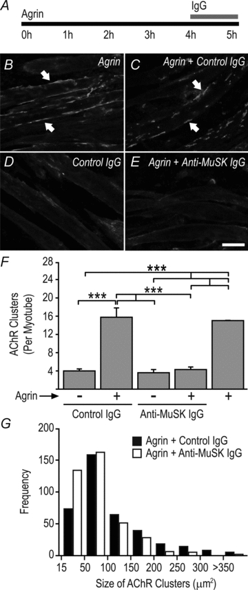
Figure 5. MuSK autoantibodies cause disassembly of pre-existing AChR clusters in culture.

A, mouse C2 myotubes were exposed to 1 nm agrin for 4 h to form large AChR clusters. Human IgG (4.5 mg ml−1 final) was then added to the culture for 1 h in the continued presence of agrin. B, agrin-treated myotubes (no IgG) displaying large AChR clusters (arrows). C, agrin treatment followed by control human IgG. D, myotubes treated with control IgG alone (no agrin). E, agrin treatment followed by exposure to MuSK2 IgG for 1 h. Scale bar is 25 μm. F, counts of the number of AChR clusters per myotube segment. All AChR clusters larger than 15 μm2 were counted. Bars show mean +s.e.m. for n= 3 culture experiments. G, frequency distributions representing the size of AChR clusters from myotubes treated with agrin followed by either MuSK2 IgG or control IgG for 1 h.
