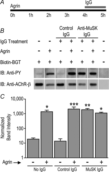
Figure 7. MuSK autoantibodies increased tyrosine phosphorylation of the AChR β-subunit.

A, myotubes were exposed to neural agrin (1 nm) for 4 h followed by human IgG (4.5 mg ml−1) for a further 1 h in the continued presence of agrin. B, sample immunoblot showing bands labelled with anti-phosphotyrosine (anti-PY; upper panel) and re-probed with anti-AChR-β-subunit (mab124; lower panel). The last lane is a control for non-specific precipitation (biotin–α-bungarotoxin was omitted). C, quantification of AChR β-subunit phosphorylation. Bars represent the mean +s.e.m. of n= 3 culture experiments. Asterisks indicate bars that were significantly different from the far left control bar.
