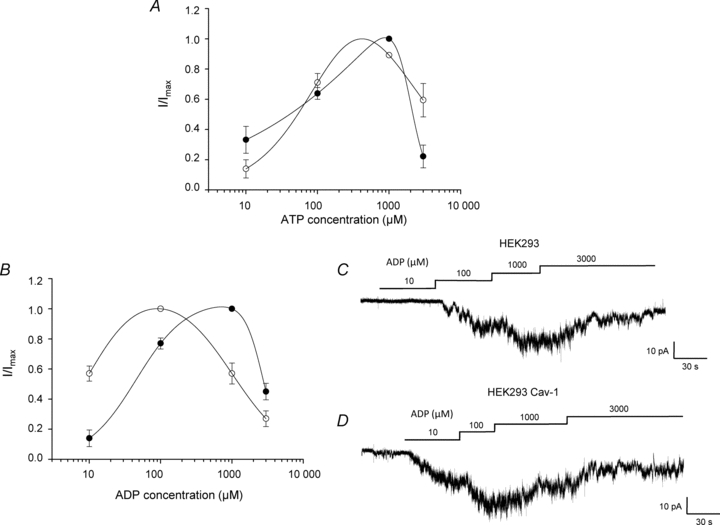
Figure 6. Caveolin alters vascular KATP channel sensitivity to MgADP.

A and B, concentration–response relationship of the ATP-induced (A) or ADP-induced (B) activation of recombinant Kir6.1/SUR2B channels in the presence 10 μm pinacidil for both caveolin-null HEK293 (filled symbols) and HEK293 caveolin-1 (open symbols) cells (means ±s.e.m., n= 6, 6 patches respectively). Channels were recorded in inside out patches at −60 mV. Data are fitted with a biphasic concentration–effect curve (see eqn (1); Methods). C and D, ADP-induced concentration-dependent activation of recombinant vascular KATP channels in inside-out patches pulled from caveolin-null HEK293 cells (C) and HEK293 cells stably expressing caveolin-1 (D).
