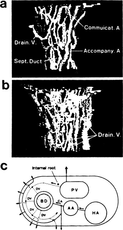
Fig 1.
Computer-aided 3-D pictures of normal peribiliary microvasculature showing (a) the afferent system; (b) the draining system; (c) a schema showing the basic pattern of periductal vasculature. BD, bile duct; AA, accompanying artery; CA, communicating arteriole; HA, larger hepatic artery; PV, portal vein; DV, draining venule.

