Fig 3.

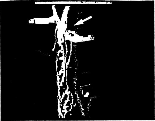
A 3-D picture of peribiliary vessels from a failed graft (Case 1). The interlobular duct has undergone marked atrophy in the upper ⅓, where CAs are destroyed and periductal capillaries are mostly missing.

A 3-D picture of peribiliary vessels from a failed graft (Case 1). The interlobular duct has undergone marked atrophy in the upper ⅓, where CAs are destroyed and periductal capillaries are mostly missing.