Figure 7.
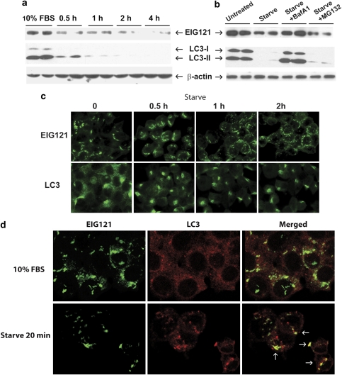
After starvation, EIG121 and LC3 colocalize and both are degraded by a lysosomal mechanism. (a) MCF-7 cells were starved in HBSS for 0, 0.5, 1, 2, and 4 h. Note the rapid degradation of EIG121 and both LC3-I and LC3-II on starvation. This experiment was conducted twice and each time with all the treatment groups in duplicate. (b) The starvation-induced degradation of EIG121 and LC3 is blocked by lysosomal inhibitor BafA1. MCF-7 cells were pretreated with either BafA1 (100 nM) or MG132 (10 μM) for 30 min, before being starved in HBSS for 2 h in the continuous presence of BafA1 or MG132. Either 50 or 150 μg of cell lysates was resolved by SDS-PAGE gel and probed by EIG121 or LC3 antibodies, respectively. (c) Immunofluorescence staining of EIG121 and LC3 at different times after starvation. A rabbit polyclonal antibody against LC3 was used. Note the scattered vesicular staining of EIG121 after starvation. (d) EIG121 and LC3 double labeling of MCF-7 cells cultured in 10% FBS or starved in HBSS for 20 min. In this experiment, a mouse monoclonal antibody against LC3 was used. The arrows indicate colocalized LC3- and EIG121-positive vesicles

