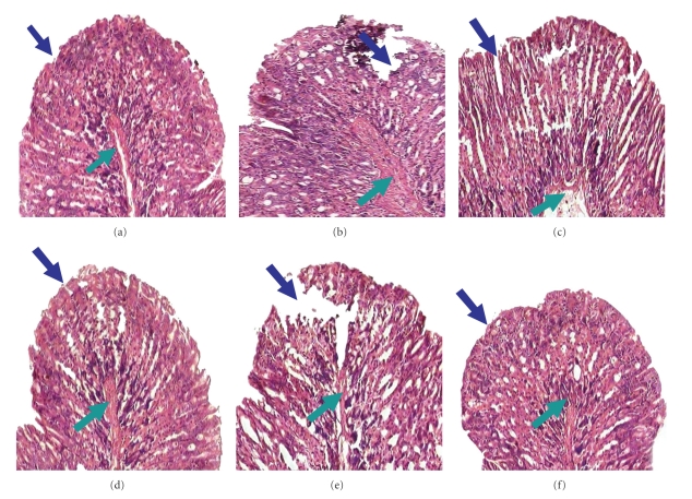
Figure 1.
Histology of mouse gastric tissue after ulcer induction by indomethacin and the effect of eAE. Ulceration in mice was induced by indomethacin (18 mg /kg, p.o.). eAE at different doses (40 mg/kg, 60 mg /kg, and 120 mg/kg) and Omeprazole (3 mg/kg) were administered 6 h post ulcer induction as described in Section 2. At the third day of ulceration, mice were sacrificed, and the stomachs were sectioned for the histological studies. Histological photograph of sham-treated (a), ulcerated untreated (b), eAE-(40 mg /kg) treated (c), eAE-(60 mg /kg) treated (d), eAE-(120 mg /kg) treated (e), and omeprazole-(f) treated mice stomach section stained with hematoxylin and eosin, present here. Gastric tissue sections were photographed at a 40X magnification. Mucosal and submucosal layers are shown by blue and green arrows, respectively.

