FIG. 4.
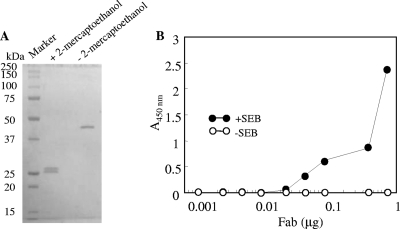
Characterization of purified Fab fragment binding to SEB. (A) SDS-PAGE of the purified Fab fragment. Purified Fab fragment (1 μg) was subjected to SDS-PAGE under nonreducing or reducing conditions and stained by Coomassie blue staining. Left lane: molecular mass marker proteins. (B) Serially diluted Fab fragment was poured onto SEB-coated (•) or uncoated (○) polystylene plates. After washing, the plate Fab fragment was detected by using HRP-labeled protein L.

