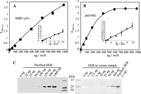
FIG. 8.
Application of MBP-scFv by ELISA and Western blotting. (A and B) Detection of SEB by ELISA. MBP-scFv (A) or ab53981 (B) was poured onto an SEB-coated polystyrene plate. After the plate was washed with buffer, SEB was detected by using HRP-labeled anti-MBP monoclonal antibody (A) or HRP-labeled anti-mouse IgG polyclonal antibody (B). Color development was done for 5 and 30 min using 50 to 1,000 ng of SEB and 0.5 to 10 ng of SEB, respectively. (C) Western blot detection of SEB by MBP-scFv. SEB solutions diluted with TBS (left) or serum (right) were separated by SDS-PAGE and subjected to Western blot analysis with MBP-scFv antibody for primary detection.

