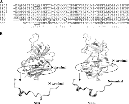
FIG. 9.
N-terminal region of SEB recognized by Fab fragment and scFv. (A) The amino acid sequences of SEs were aligned by using CLUSTAL W software. The conserved LHK motif is underlined. (B) Comparison of the three-dimensional structure between SEB and SEC2 (PDB ID; 1STE) at the N-terminal 20-residue region. The short α-helix structures of SEB and SEC2 are indicated by a bold black line.

