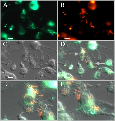
FIG. 4.
Dual infection of RAW 264.7 macrophages by differentially labeled (green and red) B. pseudomallei bacteria. Infections were carried out identically to those described in the legend of Fig. 3. (A) The green fluorescent signal indicates where gfp-tagged B. pseudomallei bacteria are replicating inside macrophages. (B) The red fluorescent signal was obtained from the same field and shows where rfp-tagged B. pseudomallei bacteria are replicating within macrophages. (C) A DIC image was then captured and is presented. (D) Overlay of images captured sequentially in panels A, B, and C. Images were superimposed at the time of capture using Zeiss AxioVision software. (E and F) Close-ups of the two macrophages indicated by arrows in panel D, where the two differently fluorescing B. pseudomallei strains are clearly visible and distinguishable within the macrophages and even within the same host cell. The total magnification in panels A to D is ×630, and all scale bars equal 10 μm.

