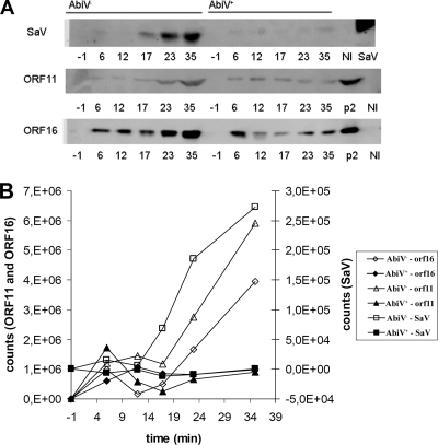
FIG. 3.
Western blotting for detection of ORF26 (SaV), ORF11, and ORF16 during phage infection. (A) Temporal phage protein production in AbiV− (left) and AbiV+ (right) L. lactis cells. Numbers represent minutes in relation to time of infection (time zero). Lanes NI, noninfected cells; lane SaV, purified SaV; lanes p2, CsCl-purified p2 virions. (B) Graphical representation of the expression levels shown in panel A. Open symbols represent AbiV−, while closed symbols represent AbiV+.

