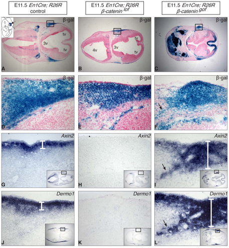
Fig. 4.
Wnt signaling is necessary and sufficient for dermal specification of cranial dermis at E11.5. Comparison between En1Cre; R26R control (β-cateninflox/+), loss-of-function (β-cateninflox/null) and gain-of-function (β-cateninflox3/+) mutant embryos. (A-F) X-gal stained transverse sections of the supraorbital region at E11.5 (G-I) Section in situ hybridization of Wnt-responsive Axin2 mRNA and (J-L) the dermal progenitor marker (Dermo1) mRNA expression. Wnt signaling activity levels affect Axin2 and Dermo1 mRNA expression. In the gain-of-function mutants, there were both ectopic (arrows in F, I, L) and expanded domains of En1CreRR, Axin2 and Dermo1+ cells (compare white brackets in G and J with I and L). 3v, third ventricle; 4v, fourth ventricle; tv, telencephalic vesicle; lof, loss-of-function; gof, gain-of-function.

