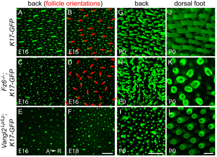
Fig. 1.
Hair follicle orientations on the back and on the dorsal surface of the feet in late embryonic and neonatal wild-type, Fz6–/– and Vangl2Lp/Lp mice. Follicles in z-stacked confocal images of skin flat-mounts are visualized by the fluorescence of a K17-GFP transgene (green). (A-I) Images of back skin are oriented with anterior to the left and posterior to the right, as indicated in E. Red arrows in B and D indicate follicle orientations for the guard hairs in the wild-type (WT) and Fz6–/– images A and C. (J-L) Images of dorsal foot skin are oriented with proximal to the left and distal to the right. Scale bars: 400 μm in A-I; 100 μm in J-L.

