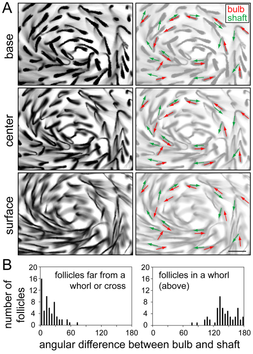
Fig. 4.
Bending of hair follicles near the center of a whorl. The whorl is from the back of the P8 Fz6–/–;Vangl2Lp/+ mouse skin shown in Fig. 6. (A) The center of the whorl is shown at three focal planes: the plane deepest within the dermis (base) shows the follicle bulbs and the plane at the epidermal surface (surface) shows the shafts as they emerge from the skin. The same three panels are reproduced on the right with the vectors for bulb (red) and shaft (green) orientations shown for 14 follicles. (B) Histograms showing bulb versus shaft angle differences for follicles from the whorl shown in A (right), and from a region of the same skin located far from a whorl or cross (left). Scale bar: 100 μm.

