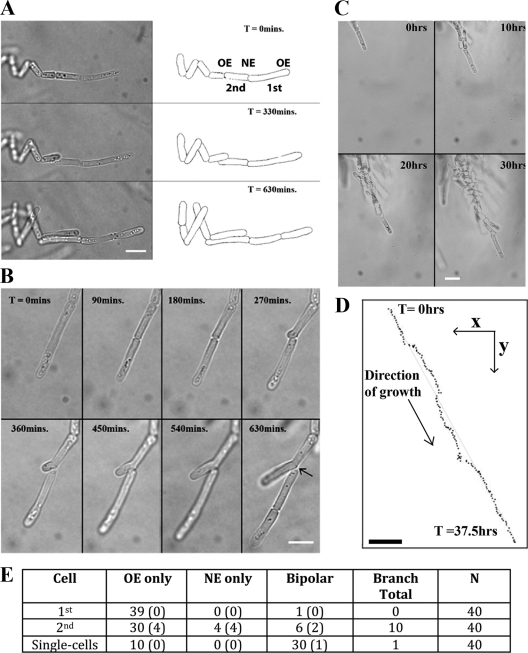
Fig. 3.
Time-lapse microscopy of S. pombe filamentous growth. (A) Montage of time-lapse images with cell outlines traced on the right. Filament tip cells are elongated (Table 2), grow monopolarly throughout the cell cycle, and undergo complete cell division, and no structures that attach adjacent cells are obvious. “1st” and “2nd” refer to the cell at the tip of the filament and the cell directly behind it, and “OE” and “NE” refer to the old and new ends, respectively. (B) Branch formation. Following division, the daughter behind the filament tip can grow monopolarly from its “new” end. More frequently, these daughters grow monopolarly from the “old” end as in panel A. (C) Selected time points from the time-lapse microscopy in Movie S1 in the supplemental material. (D) The position of the filament at each 15-min time point was plotted from Movie S1 in the supplemental material. Through 37.5 h of growth, encompassing nine cell cycles, the filament maintains a stable direction of growth. The straight guidance line represents a distance of 79.5 μm. Bar, 10 μm. (E) Quantitation of the patterns of filamentous growth. From time-lapse microscopy of growing filaments, the 1st and 2nd cells were scored as growing monopolarly from the new or old ends or bipolarly from both. The number of branched cells (branched cells shown in panel B) that were found in each class is shown in parentheses. In some cases, adjacent cells separated to allow them to grow past each other (middle image in panel A). This was classed as bipolar growth without branching. Single cells growing on LNB medium were analyzed in parallel. Time (T) is shown in minutes or hours.

