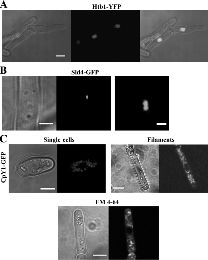
Fig. 4.
Cellular organelles in filamentous cells. (A) Filamentous cells stained with nuclear marker Htb1-YFP (YFP stands for yellow fluorescent protein) (strain JA1551). The right panel shows the transmission and fluorescence microscopy images superimposed. The extended filaments are mononucleate. Bar, 5 μm. (B) Filamentous cells stained with Sid4-GFP, a marker for the spindle pole body (SPB) (strain JA1576). The right panel is a higher magnification of the duplicated SPB seen in the left panel. Bars, 2 μm (left panel) and 0.5 μm (right panel). (C) Vacuole morphology in filamentous cells. Filamentous cells were stained with vacuole lumen marker CpY-GFP (strain JA1582) (top) or with vital dye FM 4-64 (bottom), which labels endocytic structures and vacuole membranes. The size of vacuoles appears more irregular in filaments, and they are absent from a region near the tip. All images are single planes. Bars, 5 μm.

