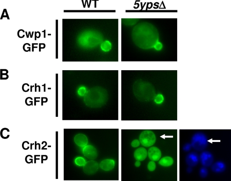
Fig. 1.
Crh2-GFP is mislocalized in the 5ypsΔ quintuple yapsin mutant. wt and 5ypsΔ cells expressing the indicated GFP fusion proteins from multicopy plasmids under endogenous promoters were grown to logarithmic phase in synthetic dextrose medium lacking uracil, harvested, and analyzed by fluorescence microscopy as described in Materials and Methods. (A) CWP1-GFP. (B) CRH1-GFP. (C) CRH2-GFP. In C, the cells were also stained with the vacuolar marker CellTracker Blue (Invitrogen) (26). Arrows indicate areas where the GFP and vacuolar stainings do not colocalize.

