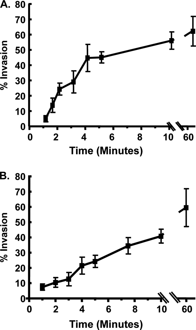
Fig. 1.

Invasion kinetics. (A) GFP+ parasites were added to HFFs in high-K+ buffer, and 20 min later invasion buffer was added to stimulate invasion. At the indicated times, the cells were fixed, and nonpermeabilized cells were stained with anti-SAG1 antibody to discriminate between extracellular and intracellular parasites. The averages and standard deviations of three independent experiments counting a minimum of 200 randomly selected parasites for each sample are shown. (B) GFP+ parasites were added to HFFs at room temperature and centrifuged for 3 min at 250 × g. The cells were then transferred to a 37°C heat block to stimulate invasion. At the indicated times, cells were fixed and then stained with anti-SAG1 to discriminate between extracellular and intracellular parasites.The averages and standard deviations of three independent experiments counting a minimum of 200 randomly selected parasites for each sample are shown.
