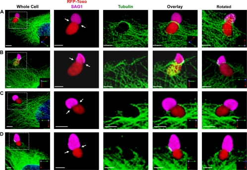
Fig. 3.
Host microtubules localize to the moving junction during Toxoplasma invasion. GFP-tubulin cells were infected with tRFP+ parasites using the synchronized invasion assay. Two minutes later, cells were fixed and processed for confocal microscopy. The left panel of each row is a low-magnification image, and the white box indicates the zoomed-in areas shown in the other panels. The rightmost panel shows the same area rotated around the x axis to show the underside of the invading parasite. (A and B) Representative 3D reconstructions of invading parasites with host microtubules associated with the moving junction (arrows). (C and D) Representative images of invading parasites without host microtubule association. Scale bars, 3 μm.

