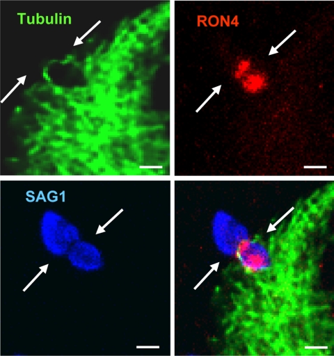
Fig. 5.
Host microtubules and RON4 colocalization at the moving junction. GFP-tubulin cells were infected with tRFP+ parasites for 2 min, methanol fixed, and then stained to detect RON4 and SAG1. The white arrows are pointing to the moving junction identified as a ring of RON4 protein at the parasite constriction (scale bar, 5 μm). The RON4 staining in front of the moving junction is apical nonexocytosed protein (1).

