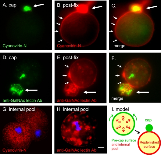
Fig. 1.
Capped cyanovirin-N-binding glycoproteins and Gal/GalNAc lectins are rapidly replenished on the E. histolytica surface from large intracellular pools. (A) Cyanovirin-N forms a tight green cap (large arrow) on the surface of trophozoites warmed for 15 min prior to fixation. (B) The same organism was fixed and then labeled with red cyanovirin-N. Cyanovirin-N binding sites that are replenished on the E. histolytica surface are marked with small arrows. (D) Similar results were obtained with a monoclonal antibody to the Gal/GalNAc lectin, which forms a tight cap on trophozoites prior to fixation (green). (E) After fixation, the Gal/GalNAc lectin that is replenished on the parasite surface is shown with a red anti-Gal/GalNAc antibody. Merged panels are as indicated. (G) Cyanovirin-N (red) binds to a large interior pool of glycoproteins in a fixed and permeabilized ameba. (H) Similar results were obtained with antibodies to the Gal/GalNAc lectin. In panels G and H nuclei are stained with DAPI (blue). Bar, 5 μm. (I) A model for the redistribution of glycoproteins binding cyanovirin-N or anti-GalNAc antibodies from large intracellular pools during capping. See Fig. S1 in the supplemental material for images of cyanovirin-N and anti-Gal/GalNAc antibody labeling of uncapped parasites. Ab, antibody.

