Fig. 3.
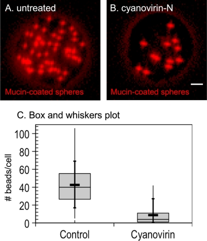
Cyanovirin-N inhibits phagocytosis of mucin-coated spheres by E. histolytica trophozoites. Fluorescence micrograph of a control E. histolytica trophozoite (A) that phagocytoses many mucin-coated spheres (red). In contrast, a representative cyanovirin-N-treated E. histolytica trophozoite (B) phagocytoses many fewer red spheres. Bar, 5 μm. (C) Phagocytosis of mucin-coated beads, as illustrated by a modified box and whiskers plot, in which the means are marked with a heavy horizontal line and the medians are marked by a light horizontal line. The lower and upper medians are the edges of each gray box, while the vertical bar shows the minimum and maximum values and the 10th and 90th percentiles (crosses).

