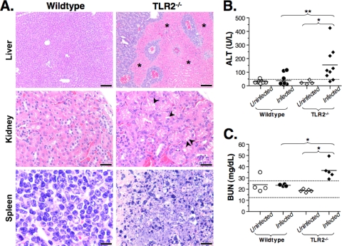
FIG. 4.
B. hermsii-infected TLR2−/− mice develop organ damage and dysfunction characteristic of septic shock. (A) Histopathological analysis was performed on hematoxylin-eosin-stained organ sections of wild-type and TLR2−/− mice at 5 days postinfection. Livers showed hypoxic foci in TLR2−/− mice (indicated with asterisks). Kidneys of TLR2−/− mice showed vacuolar degeneration (indicated with arrowheads), and the splenic white pulp showed extensive apoptosis in TLR2−/− mice. This panel reflects observations from two independent experiments involving at least five mice per group. (B and C) Serum ALT (B) or BUN (C) levels were measured in wild-type and TLR2−/− mice prior to infection and then at 4 days postinfection. The dashed line in panel B indicates the upper limit of healthy ALT levels. Dashed lines in panel C indicate the upper and lower limits of healthy mouse BUN values. These data are representative of two independent experiments. Statistically significant differences, indicated by “*” (P < 0.05) or “**” (P < 0.01) were determined by the Mann-Whitney test.

